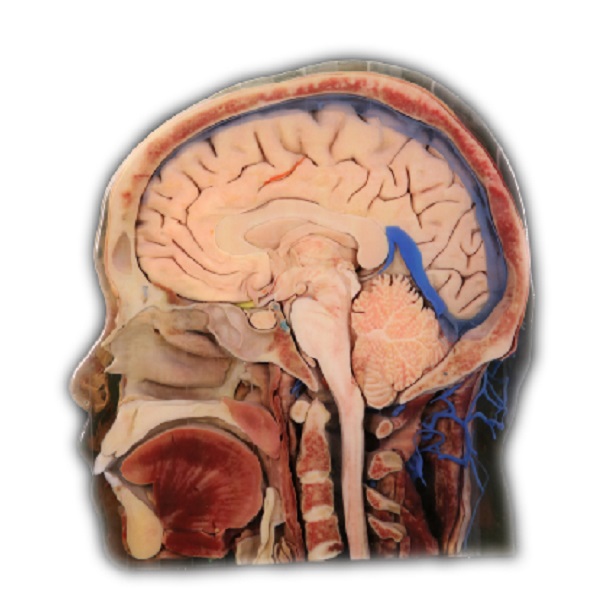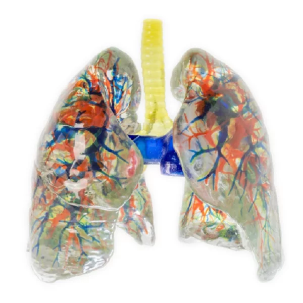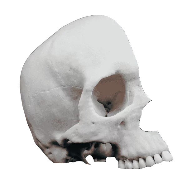
As a researcher in the field of medical education, I have come across various innovative tools that aid in enhancing learning experiences. One such tool that has gained significant attention is the anatomy bone model. These models provide a tangible representation of human bones, allowing students and professionals to study and understand the intricacies of skeletal structures.
The Benefits of Anatomy Bone Models
Anatomy bone models offer several advantages for medical education. Firstly, they provide a hands-on approach to learning, enabling individuals to physically interact with the models and gain a better understanding of anatomical structures. This tactile experience enhances retention and comprehension.
Furthermore, these models allow for detailed examination as they accurately depict the size, shape, texture, and spatial relationships between different bones. Students can explore specific features or abnormalities on these models without any time constraints or limitations.
Find more about 3d printing anatomical models.
In addition to their educational value, anatomy bone models also serve as valuable tools for surgical planning and practice. Surgeons can use these models to simulate procedures before performing them on actual patients, improving precision and reducing risks.
The Role of 3D Printing in Anatomical Models
Advancements in technology have revolutionized the creation of anatomical models through 3D printing techniques. With this technology, intricate details can be reproduced with high accuracy using various materials such as plastic or resin.
3D printing allows customization according to individual requirements by adjusting sizes or incorporating specific pathologies into the model itself. This flexibility enables educators to tailor their teaching methods based on diverse learning needs while providing an accurate representation of real-life scenarios.
Moreover, 3D printed anatomical models are cost-effective compared to traditional manufacturing methods since they eliminate the need for molds or specialized equipment. This affordability makes them more accessible to educational institutions and medical facilities.
The Advancements of DIGIHUMAN
DIGIHUMAN, a cutting-edge technology developed by researchers, takes anatomical models to the next level. It combines 3D printing with virtual reality (VR) capabilities, providing an immersive learning experience for students and professionals alike.
With DIGIHUMAN, users can interact with virtual representations of anatomy bone models in a simulated environment. This technology allows for dynamic exploration of structures, enabling users to dissect or manipulate the model digitally. The integration of haptic feedback further enhances the realism and engagement during these interactions.
Additionally, DIGIHUMAN offers collaborative features that facilitate remote learning and teamwork. Users can connect with peers or instructors from different locations to discuss cases or perform virtual surgeries together. This fosters knowledge sharing and expands opportunities for global collaboration in medical education.
Conclusion

Anatomy bone models have proven to be invaluable tools in medical education and training. Their tactile nature aids in comprehension while allowing for detailed examination of skeletal structures. With advancements like 3D printing and technologies such as DIGIHUMAN, these models continue to evolve, offering enhanced learning experiences that benefit both students and professionals in the field of medicine.
